Bridging the Gap Between What We Know and What We Do

By Jeffrey Lineberry, DDS

The dental profession is a commitment to lifelong learning. After years of school and finally graduating, many dental professionals begin practicing in the “real world”, where they quickly learn that what they learned in dental school or even in residency only scratches the surface of the many challenges in dentistry. These include patient management, clinical skills and assessment, restorative materials, technology, techniques, practice management, team management, specialist communication and support, and the list goes on. Needless to say, dental professionals have to continually grow and learn as a commitment to excellent patient care and in order to be able to stay abreast of the ever-evolving field of dentistry.

As the population ages, patients are retaining their dentitions longer and many patients are missing multiple teeth and/or have a plethora of dental issues. Many of these patients have the financial means and desires to be able to eat, smile and function properly and are now presenting to offices looking for solutions to their complex dental situations.

One of the best “teachers” in dentistry is when patient and/or clinical situations arise that challenges the dental professionals’ abilities to be able to properly diagnose and manage the patient, especially ones that have more complex situations. It is at these times the dentist can choose to either refer the patient to a more experienced practitioner, or depending on the situation, to one or more specialists.

One of the key elements is that the practitioner must learn to be able to identify these situations in a timely and proper manner so that there are no adverse effects on the patient’s overall care and clinical outcomes. Ultimately, the practitioner must be able to at least identify these clinical challenges and if referring, be able to communicate effectively to the referral source. For some clinicians, they choose to further their knowledge in clinical care so that they can either participate more in their patient care directly and/or become more effective in communicating with the specialists. 

When it comes to clinical dentistry, we only “see” what we “know”. In other words, when we all first began dental school, we all learned how to diagnose gum disease and what caries/decay “looked like” and after seeing these conditions multiple times and then having a more experienced clinician confirm what we were seeing, we could now confidently “see” and diagnose caries/decay and periodontal disease more effectively. Unfortunately, dental conditions that affect our patients are not as simple as having caries and periodontal disease.  

Many patients have complex conditions including: systemic/medical issues, temporomandibular joint(TMJ) disease, sleep issues, airway problems, poor jaw relationship, multiple missing teeth,  occlusal problems, wear,  bruxism, mouth breathing, muscle pain and the list goes on.  

In dental school and even in residency, dental professionals are limited to time and experiences with the patients that they see during that time period as well as to the experiences of their supporting colleagues. 

As a dental practitioner, if you saw one or more of these conditions or issues with a patient, would you know how to properly diagnose and manage the patient and not just simply pass the patient on for someone else to “deal with”?

Patients with complex issues require dental professionals to be willing and able to learn more about a variety of patient conditions and how they can properly diagnose and manage the patient, including proper referral. This includes developing relationships with other dental professionals and specialists who can help manage and care for these patients who have complicated conditions. 

For instance, even if a dental professional has no desire to do orthodontics on their patient population and would refer the patient for orthodontic treatment, the practitioner still needs to understand the basics of malocclusion and jaw relationship issues, so they identify the condition and refer the patient appropriately along with any concerns of the patient and/or conditions and concerns the practitioner are aware of. 

Management of a patient is not simply treating that patient in and by itself and it is not just simply referring the patient out. It is the art and science of: getting to understand and know your patient’s concerns, conditions and the patient’s understanding/ownership of their condition;  determining if you and your team are the right “fit” for the patient; gathering proper records;  identifying the issues at hand; communicating with a team of professionals when needed(interdisciplinary care); and then determining who, where and how the patient will be best managed, whether with you or someone else, with the patient at the center of it all.  Dr. L.D. Pankey referred years ago to a process  that he called the cross of dentistry: Know Yourself, Know Your Work, Know Your Patient, Apply Your Knowledge. 

In summary, dental professionals have to be willing to commit to learning outside of school and their offices. This involves time, commitment and willingness to learn and be influenced by others and to become a continual student throughout their career.  In my FDC2024 courses titled: “Bridging The Gap”: Between What We Know and What We Do” and “Mastering the Examination and Treatment Planning of the Complex Restorative Patient”, I will be sharing 20 + years of clinical and educational experience working alongside some of the most influential educators in dentistry so participants can return to their offices and “see” patients and patient care on another level.   

Dr. Lineberry will speak on this topic at the 2024 Florida Dental Convention on June 20-22 in Orlando. You can find more information on his courses at www.floridadentalconvention.com.

A New Standard of Care:  Offer Ridge Preservation for Implant Treatment Planning

By Kevin R. Suzuki DMD, MS

Ridge atrophy is well documented and dramatic.  This is well illustrated in the prospective study by Schropp et al. which demonstrated changes of an edentulous site after tooth extraction over the period of a year (1).   Edentulous ridges were shown to demonstrate an average of 50% loss of buccal-lingual dimension; the majority lost during the first 3 months.  Studies by Iasella et al. also document significant changes in apical-coronal dimension of the alveolar ridge post-extraction (2).

The patient’s goals are discussed and which option(s) best suit the situation.  In many cases options to replace an edentulous area may include:  implant-supported restoration, tooth supported-fixed prosthesis, removable prosthesis, no treatment.  Many patients may desire an option to most closely replicate the form, function, and esthetic capacity of the original tooth or teeth.  An implant-supported restoration may prove to be the most desired optimal solution.  For an edentulous site where there can be a significant compromise to native tissues and inadequate alveolar dimensions (such as a missing tooth for one year or longer), alveolar changes can complicate or rule out the possibility for placement of a dental implant. 

Ridge atrophy frequently requires regeneration of oral tissues including preservation of soft and hard tissues in preparation for placement of a dental implant.  This concept, called “ridge or site preparation/augmentation” (less appropriately “socket grafting”), has been well detailed in published texts by Suzuki and Misch (3) among a growing body of literature.  Ideally this augmentation should be performed at the time of or within 8 weeks after the extraction to ensure preservation of the maximum volume of residual bony ridge.

Clinicians may predict the degree of successful defect regeneration with bone augmentation based upon the number bony walls present (which provide vascular supply and pathways for cells and biologic factors to stimulate regeneration).  A four-wall defect is the most predictable (i.e. a completely intact tooth socket) and may not require bone augmentation if allowed to heal undisturbed long enough.  One-wall or No-wall defects prove to be the most challenging scenarios providing the least number of vascular walls for reparative and regenerative potential.  Augmentation of these defects may involve simple to multimodal techniques that incorporate one or any combination of a materials including:  bone graft, cell-occlusive membrane, biologics, tacks, screws or meshwork barriers. 

Combination grafting techniques tend to be most predictable when including the principle of guided tissue regeneration by employing an absorbable or nonabsorbable membrane.  It is through this principle, well established in wound healing literature by Melcher, that regeneration of bone around compromised teeth with bony wall defects has been shown to promote reattachment of the periodontal ligament and regrowth of alveolar bone (4).  This technique is also successful for extraction sockets. 

When considering what types of graft materials are best to use for ridge preservation the clinician should consider what ideal material characteristics are most appropriate: absence any risk of immune rejection by the recipient, predictable long term preservation of the bone in the edentulous site, working ease.  It has become commonly recommended to implement the following hierarchy when considering appropriate bone augmentation materials for implant site development:

Autogenous

Human derived allograft

Bovine, Porcine or Equine xenograft

Alloplast

Barrier membranes exclude surrounding, faster growing epithelium cells from populating an extraction socket allowing bone remodeling, deposition, and maturation. The addition of bone augmentation material acts as an osteo-inductive and/or osteo-conductive scaffold.  Angiogenesis of new blood vessels into the site promote introduction of osteoprogenitor cells.  These osteoblasts and osteoclasts utilize the mineral components of the graft material to deposit organized, osseous architecture over a period of three to six months.  Angiogenesis can be enhanced with decortication of the existing bony architecture allowing enhanced bulk and density, a principle called “regional acceleratory phenomenon”.

After healing and maturation of the graft to turn over into native bone, dental implant therapy can be predictably implemented and an implant body positioned for the ideal restorative position.  Implant dentistry has moved away from the historical approach of placing an implant body where the bone is most favorable.  The paradigm has emerged where implant treatment plans should be driven from a “restoration driven” approach.  The design of implant restoration and implant body position is dictated by the ideal prosthetic position.

Due to advances in CT scanning technology, reduced costs, and improved CT scan access the osseous architecture of a proposed implant site can be pre-operatively evaluated.  Treatment plans may be determined and predict whether augmentation may be indicated in an edentulous site.   If it is identified that there is not enough bone or it is not in the correct position to place an implant it is better to rehabilitate the ideal alveolar ridge form first.  This will permit ideal implant placement in the most appropriate prosthetic position to restore without compromise or likelihood of increased stress or restorative shortcoming in the design of the implant-prosthesis system. As dental treatment plans continue to support patients’ clinical objectives for dental implants, ridge preservation and bone grafting following tooth extraction becomes a critical component of implant site preparation.  Clinical outcomes are more predictable and successful with a basic understanding of the fundamentals of bone grafting and guided bone regeneration.

Conclusions:  It is proposed to offer, as a standard of care, ridge preservation of extraction sockets and treatment planning for the option of future dental implant(s).  Sound clinical research supports these patient therapies.

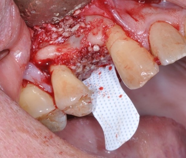
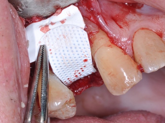
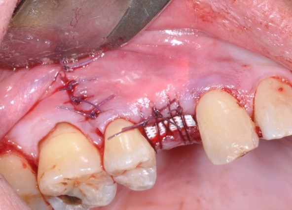
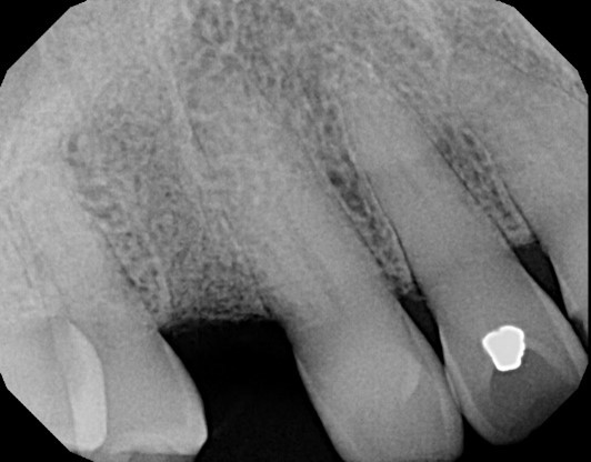
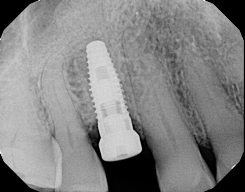
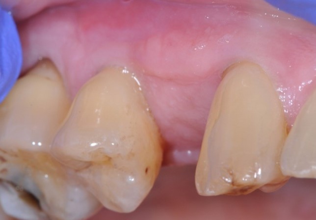
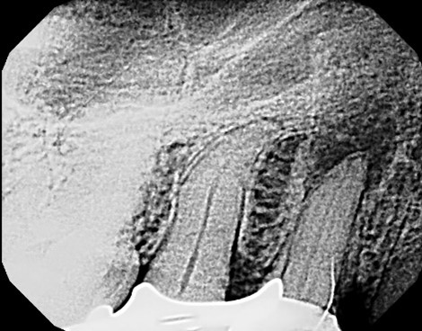
Figure Legend:  A 38 y.o. white male patient presented with a chief complaint of persisting pain in the upper right area, ( tooth #5) for days.  Diagnosis was endodontic lesion with a hopeless prognosis (Figure 1, 2). The tooth was extracted and Regional Acceleratory Phenomena performed (Figure 3), grafted with Allograft and Resorbable membrane (Figure 4 &5) employed for Guided Bone Regeneration (GBR) and sutured (Figure 6). Four months post Extraction and Ridge Preservation surgery (Figures 7 and 8, Implant was placed (Figure 9) to be restored after osseointegration period.

Acknowledgements:  contributions and support from Jon B. Suzuki DDS, PhD, MBA

Conflict of interest:  Dr. Suzuki works as a consultant for Millenium Dental Technologies

References

  1. Schropp L, Wenzel A, Kostopoulos L, Karring T. Bone healing and soft tissue contour changes following single-tooth extraction: a clinical and radiographic 12-month prospective study. Int J Perio Restor Dent. 2003; 23(4):313-23.
  2. Iasella JMGreenwell HMiller RL, et al. Ridge preservation with freeze-dried bone allograft and a collagen membrane compared to extraction alone for implant site development: a clinical and histologic study in humans. J Periodontol. 2003;74(7):990-9.
  3. Misch CE, Resnik RR, Suzuki JB, et al.  “Tooth Extraction, Socket Grafting, and Barrier Membrane Bone Regeneration.” Contemporary Implant Dentistry 4th ed. St. Louis: Mosby Elsevier, 2020. 870-904.

Dr. Suzuki will speak on this topic at the 2024 Florida Dental Convention on June 20-22 in Orlando. You can find more information on his courses at www.floridadentalconvention.com.

Advanced Hygiene Therapy – Developing a Comprehensive Approach

By Dianne Watterson, MBA, RDH, GC-C

When I became a dental hygienist in 1978, I did not foresee the “love affair” that loomed in my future, a love affair in treating periodontal disease.  Nothing gives me more professional gratification than taking a patient with frank disease to a state of good oral health through a series of appointments. 

Of course, there are many variables to consider in treatment planning comprehensive care, such as the patient’s medical history.  How might my patient with a history of chemotherapy for breast cancer respond differently than a non-chemo patient?  What outcomes should I expect and how should my care be modified for my diabetic patients?  A challenging variable is the patient’s motivation and willingness to “own” the disease and exercise behavior change.  Many patients have lifestyle factors that influence treatment outcomes, such as smoking and recreational drug or alcohol use.  What about the patient’s diet?  Can a person’s diet affect treatment outcomes?  And finally, what factors determine when the patient needs specialized care from a periodontist?  What about the patient that refuses a periodontal referral?  These are all important questions.

One of my most memorable patients was a 35-year-old patient with moderate periodontitis.  His pockets ranged from 5-7 mm, his oral hygiene was mediocre, and he was a former smoker.  He also indicated he consumed alcohol on weekends.  I completed his definitive care.  The patient was very compliant, and his oral hygiene improved significantly.  But at his first supportive therapy appointment, I noticed some of his periodontal readings had actually increased.  This was not supposed to happen.  Three months later, I knew something was not right, as his periodontal depths had not stabilized.  We referred the patient to a periodontist who asked the patient, “Tell me, how many times do you typically get up during the night to use the rest room?”  The patient replied, “Three times.”  The periodontist suspected diabetes, and a subsequent referral to the patient’s physician confirmed the diagnosis. 

My years of experience have taught me that customization is one of the most important considerations for patient success.  After all, what works for one patient may not work for a different patient.  Aspects of care that require customization include the treatment plan, the patient’s at-home oral care plan, and the supportive periodontal therapy required after the definitive care is completed.  Understanding how to communicate with clarity, compassion, and grace goes a long way in establishing connected relationships with patients.  When patients like you and sense your caring, they are more likely to comply with your instructions and recommendations for care.  When everything is finished, how do you measure success?  The measure of success comes over time.  There will be no continuing loss of supporting bone and attachment.  This outcome will not be seen in three or even six months, but in nine to twelve months following definitive treatment, the clinician will know.  To achieve this success makes the job worth all the effort!

Ms. Watterson will speak on this topic at the 2024 Florida Dental Convention on June 20-22 in Orlando. You can find more information on her courses at www.floridadentalconvention.com.
THINGS TO KNOW BEFORE YOU GO…TO FDC2023!

1. ON-SITE BADGE PICKUP HOURS & LOCATIONS

  • Wed., June 28 – 4-9 PM (Hotel Lobby)
  • Thurs., June 29 – 7 AM-8 PM (City Hall Lobby) or 4-9 PM (Hotel Lobby)
  • Fri., June 30 – 7 AM-6 PM (City Hall Lobby)
  • Sat., July 1 – 7 AM-3 PM (City Hall Lobby)
  • If you are registered as “Exhibit Hall Only,” you will pick up your badge at the first-floor rotunda outside the Exhibit Hall on Thurs., 10 AM-5 PM, Fri., 8 AM-5 PM or Sat., 8 AM-1 PM.

2. FREE OFFSITE PARKING & SHUTTLE SERVICE

  • Free parking and shuttle service to the Gaylord Palms will be available for attendees at ESPN’s Wide World of Sports (700 S. Victory Way, Reunion, FL 34747). Please allow ample time, at least one hour prior to course start, for parking and shuttle service.
  • Shuttle Hours:
    • Thurs., June 29 – 7 AM-11:30 PM
    • Fri., June 30 – 7 AM- 11 PM
    • Sat., July 1 – 7 AM-6 PM

3. FDC2023 FREE MOBILE APP

  • Download the mobile app by searching “FDC2023” in the Apple App store or Google Play one week prior to FDC. To view your personalized course schedule and course/event location you must be logged into the app with your registration ID and last name.
  • You can also download handouts, search exhibitors, view the event schedule, access the Gaylord Palms maps and more with the app! Be sure to turn on your notifications for on-site updates.

4. COURSE HANDOUTS

  • Course handouts will be available within your online registration dashboard two weeks prior to FDC.
  • In an effort to “go green,” FDC will not provide handouts onsite. Please print, download on your mobile device or view in the mobile app on-site.
  • Note: some handouts may be too large to view within the mobile app. Please download and save to your device or print ahead of time.

5. EXHIBIT HALL

  • Exhibit Hall Hours:
    • Thurs., June 29 – 11 AM-6 PM
    • Fri., June 30 – 8:45 AM-6 PM
    • Sat., July 1 – 9 AM-2 PM
  • View the current exhibitor listing and find FDC-exclusive exhibitor coupons at exhibithall.floridadentalconvention.com.

PUPPY BREAK IN THE EXHIBIT HALL

Are you feeling stressed or overwhelmed? Take a break from your busy day and visit adorable puppies at our Puppy Cuddle Break on Friday, June 30 from 3-5:45 PM in the Exhibit Hall and experience the joy and comfort of cuddling with these furry friends. Adoptions will be available for those who feel a connection.

6. SOCIAL EVENTS INCLUDED IN YOUR REGISTRATION

  • Make plans to attend the social events included in your registration*
    • Thurs., June 29
      • Welcome Cocktail Reception – 4-6 PM (a drink ticket for this event will be loaded on your badge)
      • LIVE! at FDC Party– 8-11 PM
      • Guitarist in Wreckers – 10 PM-1 AM
    • Fri., June 30
      • Alumni Receptions – 5-7 PM
      • Out of This World Party – 7:30-10:30 PM
      • The After Party – 10 PM-1 AM

* Events are not included in the free “Exhibit Hall Only” registration.

7. ATTENDEE LUNCH VOUCHERS

  • If you have purchased a Thursday morning and afternoon course or have signed up to be a speaker host, you will receive a $20 lunch voucher for Exhibit Hall concessions. Vouchers can be used in the Exhibit Hall on Thursday through Saturday, 11 AM-2 PM.
  • Your lunch voucher will be automatically loaded onto your badge. Just tap and go during checkout to use.

8. CE VERIFICATION/REPORTING

  • Per the Florida Board of Dentistry, you must be present in a course 50 of 60 minutes to receive 1 hour of CE credit. Your badge will be scanned when you enter and exit a course to calculate your hours attended.
  • Your CE certificate will be emailed to you by Monday, July 3. CE certificate printing stations will be available on-site Thursday and Friday, 9 AM-6 PM and Saturday, 9 AM-5:30 PM.
  • CE credit will be reported to CE Broker for all Florida-licensed attendees by July 29, 2023.

GET STEP-BY-STEP DIRECTIONS AT THE GAYLORD PALMS

  • Need help navigating to your FDC courses at the Gaylord Palms? Download the “Gaylord Hotels” app in the Apple App store or Google Play upon arriving at FDC.
  • Select the hotel “Gaylord Palms” and then click “Find My Way” to have the app access your location and give you step-by-step navigation to your courses.

Find out more at www.floridadentalconvention.com.